

The name may suggest it was built for missions to the Red Planet, but Christchurch-based MARS Bioimaging’s innovations are firmly grounded on Planet Earth. That being said, its imaging scanner has the potential to revolutionise medicine.
Known as the Medipix All Resolution System (MARS), the imaging machine can take multi-energy “true colour” x-ray images and analyse what is shown in the picture, such as the interior contents of a tumour.
In other words: it takes anx-ray, and can tell you what is inside the x-ray. And as for applications? Think enabling medical procedures, such as letting doctors know if it’s safe to operate on what might be a tumour on a patient’s neck close to a major artery.
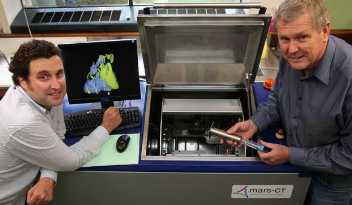
Behind MARS is the father-son duo of University of Canterbury professor Dr Phil Butler and associate professor Dr Anthony Butler. Dozens of current and completed UC PhD students have also been involved, along with masters and honours students.
As Phil Butler told Idealog, the MARS represents a significant improvement over traditional x-ray or magnetic resonance imaging (MRI), which can only analyse the shapes of objects and not the content that makes them up.
“If you’ve got an infection in a knee joint replacement, with a standard system you can’t actually see the infection. But you’ll be able to see it with this.”
Inside the scanner is a 14mm-square chip, a semiconductor that measures the particle properties of an x-ray photon, as well as its energy and position. A tissue sample can be placed inside the MARS, which takes a few minutes to create 3D images. After that, the images are then reconstructed and recorded on a computer, where they can then be accessed and analysed by whoever needs to look at them.
As Anthony Butler also told Idealog: “You can identify or characterise or measure components of a tissue [with the MARS]. You can measure the water content or the fat content or the calcium content [for example]. If you compare that to an ordinary x-ray, you can’t do that at all.”
Funding for the MARS began in 2002 with a $350,000 grant from the New Economy Research Fund. A $1.5 million grant from the Tertiary Education Fund followed, as did an additional $4.5 million from what is now known as the Ministry of Business, Innovation and Employment (MBIE) for a version that could be used on small animals. In late 2014, the MBIE poured in $12 million for development of a human-sized version. In total, the scanner has received more than $20 million in direct Government funding over more than a decade.
And that doesn’t even include the support through student scholarships, staff time and access to equipment.
Anthony Butler says that while the scanner has myriad applications, much of the research so far has been focused on medicine. And researchers from institutions including Yale and the Mayo Clinic have collaborated on MARS, while other researchers and scientists regularly visit.
“We’ve had to work more closely with the medical researchers who have brought their own skill sets. So we’ve been working with people who look at blood vessels or atheromas [when degenerative material accumulates in artery walls] or vascular disease, and particularly around strokes and the blood vessels in the neck. We’ve been working with cancer researchers, and we’ve been working with a lot with people on joint implants and metal implants for knee replacements and cartilage health. But there are dozens of other applications.”
